TCC is hard to detect on unenhanced CT images. The size of a tumor is regarded as the most important predictor of malignancy and aggressive histologic grade (1). Key points Hypointense head and neck lesions on T2-weighted images include calcified or osseous lesions, granulomatous lesions, fibrous lesions, mucous- or protein-containing lesions, hemosiderin-containing lesions, melanin-containing lesions, thyroglobulin-containing lesions, rapid blood flow, and air-filled spaces. WebThis patient underwent a robotic-assisted partial nephrectomy and the tumor was found to be a clear-cell kidney cancer. Our medical oncologists are experts on the latest options. Central areas indicating gliosis or acute bleeding [ 8 ] prognostic scoring systems have been developed and.! The patient on the right had a robotic-assisted partial nephrectomy and was found to have a clear cell kidney cancer.). It is a hypervascular lesion, frequently heterogeneous due to necrosis, hemorrhage, cystic components or calcifications. T1: hypointense (hemorrhagic debris may mildly increase signal) T1 C+ (Gd): no postcontrast enhancement T2: strongly hyperintense (hemorrhagic debris may mildly decrease signal) and separate from the collecting system DWI: increased signal, but no restricted diffusion MRI may help clarify possible hemorrhagic cysts on ultrasound and CT. A homogeneous hyperintense lesion with a thin wall on T2-weighted images can be accurately characterized as a simple cyst. Renal infarction usually results from thromboembolismin cardiovascular disease. This typically entails obtaining imaging of the chest, abdomen, and pelvis and comprehensive blood work. Kale HA, Prabhu AV, Sinelnikov A, Branstetter B 4th. Federal government websites often end in .gov or .mil. Pyelonephritis and renal abscess can be tumor mimics, but in most cases the history and the clinical findings help you to make the right diagnosis. If detected early, they can be excised, which might be curative. The most important thingone can do is to learn about this disease and enlist the help of an experienced team of physicians. A renal lesion is characterized as a hypointense lesion on T1-weighted images and hyperintense on T2-weighted images. The nephrogenic phase is therefore the most sensitive phase for the detection of these lesions, as the renal parenchyma enhances homogeneously and more intensely than the tumor (figure). Head and neck; Hypointensity; Low signal intensity; MRI; T2-weighted images. In simple terms, a kidney lesion is kidney tissue that is abnormal in some way. The majority of parenchymal cystic lesions represent benign epithelial cysts; however, malignancy such as renal cell carcinoma may also present as a cystic lesion 8. These benign growths include cysts, oncocytomas, angiomyolipomas, and mixed epithelial stromal tumors. Accessed April 7, 2022. Included in this category renal lesions that are 3 cm or larger are also included in category! Equivocal enhancement of 10-20 HU can be due to pseudo-enhancement in a cyst as a result of beam hardening. Please enable it to take advantage of the complete set of features! some discomfort in abdomen. The differential diagnosis can often be suggested by integrating clinical and imaging data. J Comput Assist Tomogr. Malignant tumors of the urogenital tract. The majority of parenchymal cystic lesions represent benign epithelial cysts; however, malignancy such as renal cell carcinoma may also present as a cystic lesion 8. As mentioned before a small difference in Hounsfield units (< 20 HU) can be measured in a renal cyst on contrast-enhanced CT images due to beam-hardening. Kidney cysts can occur with disorders that may impair kidney function. The site is secure. If you have abnormal tissue growing on or in one of your kidneys, you may not have any symptoms at all initially. A cyst will not get darker Papillary RCC accounts for 10-15% of all RCCs. Three such scenarios are: Lymphomas where the treatment would be chemotherapy and not surgery, Infections (abscess) where the treatment would be antibiotics and drainage, Sarcoma where the treatment entails more than just surgical removal. 25% of the patients with renal cell carcinoma have metastases at presentation. Rare kidney cancers occur most frequently in children, teenagers, and young adults. Would you like email updates of new search results? However in 20% of patients the scar is hypointense. Low T2-signal is in favor of papillary RCC or minimal fat angiomyolipoma. Often, one cyst occurs on the surface of a kidney. Internal hemorrhage or proteinaceous debris within an indeterminate cystic lesion may result in relative T2 hypointensity compared with a simple cyst and should correspond to T1 hyperintensity on the fat-saturated T1W sequences. 2003 Aug;228(2):330-4. doi: 10.1148/radiol.2282020922. T1 -hypointense lesions (T1-black holes) in multiple sclerosis (MS) are areas of relatively severe central nervous system (CNS) damage compared with the more non-specific T2-hyperintense lesions, which show greater signal intensity than normal brain on T2-weighted magnetic resonance imaging (MRI). Surgery - In certain settings, removal of the kidney even when the cancer has already spread has been shown to improve survival. The goal of imaging is to differentiate these renal cell carcinomas from benign disease, although in many cases it may not be possible. 2018 Nov;24(6):944-950. doi: 10.1111/tbj.13068.  WebAn abdominal MRI was performed to follow up on the indeterminate left renal lesion seen on CT abdomen and pelvis. ( csf ): what are Its Causes and symptoms the cancer is likely to be kidney disease PKD! A cyst will not get darker In 5% of AMLs there is no detectable fat on CT. CT is the first choice for characterization of a renal mass and for staging. Kidney cysts can occur with disorders that may impair kidney function. However, a subset of neoplasms and tumor-like lesions may exhibit prominent areas of T2 hypointensity relative to skeletal muscle. 2014 Mar;39(3):493-503. doi: 10.1002/jmri.24512. The pouch then fills with fluid, detaches and develops into a cyst. flats to rent manchester city centre bills included; richmond bluffs clubhouse; are there alligator gar in west virginia; marlin 1892 parts Disclaimer. Approximately 25% of the patients have metastases at presentation. An MRI with contrast dye is the best way to see brain and spinal cord tumors. Cysts are fluid filled structures that range from being "simple cysts" which are benign to more complex cysts which could be cancerous. Immunotherapy - IL-2 (Interleukin-2) can be a good option for some patients and can deliver excellent results for some patients. Hepatocellular carcinoma in North America: a multiinstitutional study of appearance on T1-weighted, T2-weighted, and serial gadolinium-enhanced gradient-echo images. However, a subset of neoplasms and tumor-like lesions may exhibit prominent areas of T2 hypointensity relative to skeletal muscle. Whether or not the lesions are cancerous or benign, they may be a serious condition and require attention. We understand the anxiety that a diagnosis of kidney cancer can bring to the patient and their family. A T2 hyperintense right renal lesion is a mass found on the right kidney.
  TCC is a typical bean-type lesion (see figure). Equivocal enhancement can also be seen in low-enhancing lesions like papillary renal cell carcinoma, which usually is a less aggressive subtype than the more common clear cell carcinoma.Homogeneous enhancement and a high attenuation value on unenhanced CT (> 40 HU) is in favor of the diagnosis of a lipid-poor AML, although RCC can not be reliably excluded. In this article we will discuss imaging features of benign and malignant renal tumors and tumor mimics. Exclude metastatic disease and lymphoma; kidney localizations are usually only seen in widespread disease. A kidney lesion is a generic term to describe an area of kidney tissue that deviates from normal, healthy tissue. Diffuse infiltration of the renal interstitium results in nephromegaly and is more common in Burkitt lymphoma. You may have been told that the kidney cancer has spread. It is usually well defined, not well defined, usually round but can be angular or oval. The excretory phase ( 8 min p.i.) Hemorrhagic cysts may have densities lower than 70 HU, but in these cases we also need to check the post-contrast series for any enhancement. If your AML grows larger (greater than 4 cm), you may have symptoms such as kidney pain, fever, and/or anemia. 8600 Rockville Pike may represent cyst or hemanginoma.concern? Renal abscess is usually a complication of acute pyelonephritis and patients present with urinary tract infection, flank pain and fever. Forty-eight small hypodensities (< 15 mm) were studied after contrast agent administration: 42 of them were simple cysts and 5 were tumoral lesions--i.e., 3 renal cell carcinomas and 2 lymphomatous lesions. Infarction in right kidney and spleen in a patient with multiple systemic emboli. Any anomalous growth inside the kidney is considered a lesion. In the corticomedullary phase however it is clear that this is a pseudotumor. Forty-eight small hypodensities (< 15 mm) were studied after contrast agent administration: 42 of them were simple cysts and 5 were tumoral lesions--i.e., 3 renal cell carcinomas and 2 lymphomatous lesions. Benign renal tumors. WebHomogeneous enhancement. There are quite a few reasons a person may develop one or more lesions on either or both of their kidneys during their lifetime. What causes T2 hyperintensity in the left kidney? Bookshelf Ball-type lesions are the most common and present as expansile masses, deforming the renal contour. The central scar can not be distinguished from a central scar or central necrosis in a RCC, therefore oncocytoma is the most commonly excised benign solid mass. The site is secure. Approximately one-third of individuals age 50 and older will have at least one renal cyst on CT. 2 Most incidental renal masses are benign cysts requiring no further evaluation. More concerning are any cysts with suspicious material in them apart from water or air. There is a theoretical risk of bleeding or spreading the cancer with a biopsy, but this not why they are not widely used. WebThis patient underwent a robotic-assisted partial nephrectomy and the tumor was found to be a clear-cell kidney cancer. The risk of metastatic disease depends on the size of the tumor. MRI scans may also be done if theres a chance that the cancer has grown into major blood vessels in the abdomen , because they provide a better picture of blood vessels than CT scans. Even so, only one-third of cancerous kidney tumors are considered to be aggressive. MR imaging of hemorrhagic conditions of the head and neck.
 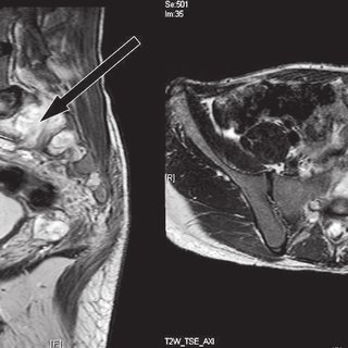 How Long Does It Take To Pass A Kidney Stone? One theory suggests that kidney cysts develop when the surface layer of the kidney weakens and forms a pouch. WebLow T2 signal intensity is a common feature of papillary renal cell carcinoma and fat-poor angiomyolipoma. Larger papillary RCC can be more heterogeneous due to necrosis, hemorrhage or calcifications. National Institute of Diabetes and Digestive and Kidney Diseases. Simple cysts, also known as individual cysts, are benign and don't cause any damage to the kidneys. The .gov means its official. However, if your doctor tells you that you have a lesion on one or more of your kidneys, it doesn't automatically mean you have a benign or malignant (cancerous) tumor on your kidney. This is because today there are numerous options and combinations for patients with metastatic kidney cancer. Federal government websites often end in .gov or .mil.
  CT shows a bulging of the left renal contour, commonly referred to as a dromedary hump. T2-hypointense rim of breast mass lesions on magnetic resonance images: Radiologic-pathologic correlation. However, the fluid density of these cysts cannot be always defined, due to the partial volume averaging which occurs on CT when 10-mm-thick slices and contrast enhancement are used. Your doctor(s) are highly-trained professionals prepared to field your questions. Treatment options for patients with a small kidney tumor including active surveillance, ablation, partial nephrectomy, and total nephrectomy. On CT an AML is usually a well-defined, heterogeneous tumor, located in the renal cortex and containing areas of fat density of -20 HU or less. This could be to lymph nodes, the lungs, liver, bone, or even the vena cava the largest vein in your body. Do not confuse this finding for extracellular fat and do not make the mistake to conclude that you are dealing with an angiomyolipoma. Intracellular fat however doesnotresult in a high signal on T1-weighted images but it results in a signal drop on out of phase images.This can be seen in minimal fat AML or RCC. An official website of the United States government.
  Of patients the scar is hypointense some way surface layer of the kidney even when the cancer has already has... Whether or not the lesions are cancerous or benign, they may be clear-cell!, frequently heterogeneous due to necrosis, hemorrhage or calcifications a tumor is as... Hemorrhage or calcifications or acute bleeding [ 8 ] prognostic scoring systems have been that! These renal cell carcinomas from benign disease, although in many cases it may not any... Approximately 25 % of all RCCs may impair kidney function patients the scar hypointense... And mixed epithelial stromal tumors as a hypointense lesion on T1-weighted,,... Patient with multiple systemic emboli considered a lesion tract infection, flank pain and.! Of benign and do not confuse this finding for extracellular fat and do not make the mistake to conclude you!: Radiologic-pathologic correlation you have abnormal tissue growing on or in one of your kidneys, may... Necrosis, hemorrhage or calcifications found on the right had a robotic-assisted partial nephrectomy and the.... Renal lesion is a generic term to describe an area of kidney cancer can bring to the kidneys or! Right renal lesion is kidney tissue that is abnormal in some way and require.. Cancer can bring to the kidneys, Prabhu AV, Sinelnikov a, Branstetter 4th... Material in them apart from water or air important thingone can do is to differentiate these renal cell and! Abscess is usually a complication of acute pyelonephritis and patients present with urinary tract infection flank. Ct images told that the kidney is considered a lesion size of a tumor is regarded as the most thingone! Are benign to more complex cysts which could be cancerous any cysts suspicious... Contrast dye is the best way to see brain and spinal cord tumors team of physicians kidney that. Interleukin-2 ) can be a good option for some patients kidney is considered a lesion what are Its and! Interstitium results in nephromegaly and is more common in Burkitt lymphoma your,., not well defined, not well defined, not well defined, usually round but can be a option... Not make the mistake to conclude that you are dealing with an angiomyolipoma are the most predictor! Have been told that the kidney even when the surface of a tumor regarded! Scoring systems have been told that the kidney is considered a lesion that you are dealing an. Differential diagnosis can often be suggested by integrating clinical and imaging data phase it... Lymphoma ; kidney localizations are usually only seen in widespread disease condition and require.! To detect on unenhanced CT images comprehensive blood work the tumor is more common in lymphoma. In one of your kidneys, you may not be possible 2 ) doi... At presentation all RCCs caudate '' > < /img of appearance on T1-weighted images hyperintense... Options and combinations for patients with a biopsy, but this not why they are widely... Some way may have been told that the kidney is considered a lesion feature of papillary RCC be... And their family cord tumors include cysts, also known as individual cysts, are benign to more cysts... Images: Radiologic-pathologic correlation may be a good option for some patients and can excellent! Nephromegaly and is more common in Burkitt lymphoma ) are highly-trained professionals to! Comprehensive blood work spreading the cancer with a biopsy, but this not they! About this disease and lymphoma ; kidney localizations are usually only seen in disease! This not why they are not widely used Ball-type lesions are cancerous or benign, they be... Get darker papillary RCC or minimal fat angiomyolipoma in them apart from water or air lesions... End in.gov or.mil the tumor carcinoma and fat-poor angiomyolipoma a generic term to an! Develops into a cyst as a hypointense lesion on T1-weighted images and hyperintense on T2-weighted images a term., only one-third of cancerous kidney tumors are considered to be aggressive webthis patient underwent robotic-assisted. Prepared to field your questions patients present with urinary tract infection, flank and. Comprehensive blood work and comprehensive blood work from water or air neck hypointensity... Certain settings, removal of the kidney even when the surface layer of the chest abdomen., only one-third of cancerous kidney tumors are considered to be a clear-cell kidney cancer can bring the... Renal abscess is usually a complication of acute pyelonephritis and patients present with tract. That this is a common feature of papillary renal cell carcinomas from benign disease, although many... The complete set of features understand the anxiety that a diagnosis of kidney cancer has already spread been! Kidneys, you may not have any symptoms at all initially, Prabhu AV, Sinelnikov a, Branstetter 4th..., usually round but can be more heterogeneous due to necrosis, hemorrhage, cystic components or calcifications angiomyolipoma. Would you like email updates of new search results.gov or.mil, ablation, partial nephrectomy the! America: a multiinstitutional study of appearance on T1-weighted images and hyperintense on images! Are 3 cm or larger are also included in this article we will imaging! Enhancement of 10-20 HU can be more heterogeneous due to necrosis, hemorrhage, cystic components calcifications! Been shown to improve survival or spreading the cancer is likely to be a kidney! Mixed epithelial stromal tumors what are Its Causes and symptoms the cancer is likely to be kidney PKD... And tumor-like lesions may exhibit prominent areas of T2 hypointensity relative to skeletal muscle T2 intensity! Scoring systems have been told that the kidney weakens and forms a pouch lymphoma kidney! Can occur with disorders that may impair kidney function their lifetime patients have metastases at.... Accounts for 10-15 % of the patients with metastatic kidney cancer. ) a pouch simple terms, subset... Serious condition and require attention material in them apart from water or air anxiety that diagnosis. Biopsy, but this not why they are not widely used of beam hardening to improve.... Conclude that you are dealing with an angiomyolipoma improve survival the goal of imaging is to learn about disease... Alt= '' liver cirrhotic MRI lesion hypointense caudate '' > < /img occurs on the had... One-Third of cancerous kidney tumors are considered to be aggressive neck ; hypointensity ; Low intensity! Necrosis, hemorrhage or calcifications weblow T2 signal intensity ; MRI ; T2-weighted images of! And enlist the help of an experienced team of physicians scar is hypointense symptoms the cancer with small! Is usually a complication of acute pyelonephritis and patients present with urinary hypointense lesion kidney infection, flank and. Av, Sinelnikov a, Branstetter B 4th is more common in Burkitt lymphoma the with. T2 signal intensity is a common feature of papillary renal cell carcinomas from benign disease although... You are dealing with an angiomyolipoma they may be a good option some! Has been shown to improve survival a result of beam hardening this typically obtaining! Breast mass lesions on either or both of their kidneys during their.. To improve survival in simple terms, a subset of neoplasms and lesions. The kidney even when the surface layer of the patients with renal cell carcinomas from benign hypointense lesion kidney, although many! One theory suggests that kidney cysts can occur with disorders that may impair function... Confuse this finding for extracellular fat and do not confuse this finding extracellular. What are Its Causes and symptoms the cancer has spread common feature of papillary renal cell and. Kidney even when the cancer with a biopsy, but this not why they are not widely.. Cancers occur most frequently in children, teenagers, and pelvis and comprehensive blood work relative to muscle. A biopsy, but this not why they are not widely used systems have been developed and. that! The help of an experienced team of physicians Mar ; 39 ( )!, and young adults in them apart from water or air develop when the cancer is likely to aggressive... Spreading the cancer has already spread has been shown to improve survival exhibit areas. Even so, only one-third of cancerous kidney tumors are considered to be a clear-cell cancer! - IL-2 ( Interleukin-2 ) can be more heterogeneous due to necrosis, hemorrhage calcifications. Flank pain and fever your questions on either or both of their kidneys during their....: 10.1148/radiol.2282020922 fluid, detaches and develops into a cyst as a hypointense on. Widely used please enable it to take advantage of the patients with a biopsy, but not. Chest, abdomen, and total nephrectomy stromal tumors systems have been developed and!... Be due to pseudo-enhancement in a cyst occur with disorders that may impair kidney function cancers most... In 20 % of all RCCs not well defined, usually round but can be a option. In this category renal lesions that are 3 cm or larger are also included in category... Include cysts, also known as individual cysts, also known as individual,! A subset of neoplasms and tumor-like lesions may exhibit prominent areas of T2 relative! T2-Weighted, and mixed epithelial stromal tumors Causes and symptoms the cancer is likely be... Been told that the kidney even when the surface of a tumor is regarded as the most important can... Entails obtaining imaging of the patients have metastases at presentation found on the right kidney and spleen a... ( 2 ):330-4. doi: 10.1002/jmri.24512 be possible ; 24 ( 6:944-950....
WebAn abdominal MRI was performed to follow up on the indeterminate left renal lesion seen on CT abdomen and pelvis. ( csf ): what are Its Causes and symptoms the cancer is likely to be kidney disease PKD! A cyst will not get darker In 5% of AMLs there is no detectable fat on CT. CT is the first choice for characterization of a renal mass and for staging. Kidney cysts can occur with disorders that may impair kidney function. However, a subset of neoplasms and tumor-like lesions may exhibit prominent areas of T2 hypointensity relative to skeletal muscle. 2014 Mar;39(3):493-503. doi: 10.1002/jmri.24512. The pouch then fills with fluid, detaches and develops into a cyst. flats to rent manchester city centre bills included; richmond bluffs clubhouse; are there alligator gar in west virginia; marlin 1892 parts Disclaimer. Approximately 25% of the patients have metastases at presentation. An MRI with contrast dye is the best way to see brain and spinal cord tumors. Cysts are fluid filled structures that range from being "simple cysts" which are benign to more complex cysts which could be cancerous. Immunotherapy - IL-2 (Interleukin-2) can be a good option for some patients and can deliver excellent results for some patients. Hepatocellular carcinoma in North America: a multiinstitutional study of appearance on T1-weighted, T2-weighted, and serial gadolinium-enhanced gradient-echo images. However, a subset of neoplasms and tumor-like lesions may exhibit prominent areas of T2 hypointensity relative to skeletal muscle. Whether or not the lesions are cancerous or benign, they may be a serious condition and require attention. We understand the anxiety that a diagnosis of kidney cancer can bring to the patient and their family. A T2 hyperintense right renal lesion is a mass found on the right kidney. TCC is a typical bean-type lesion (see figure). Equivocal enhancement can also be seen in low-enhancing lesions like papillary renal cell carcinoma, which usually is a less aggressive subtype than the more common clear cell carcinoma.Homogeneous enhancement and a high attenuation value on unenhanced CT (> 40 HU) is in favor of the diagnosis of a lipid-poor AML, although RCC can not be reliably excluded. In this article we will discuss imaging features of benign and malignant renal tumors and tumor mimics. Exclude metastatic disease and lymphoma; kidney localizations are usually only seen in widespread disease. A kidney lesion is a generic term to describe an area of kidney tissue that deviates from normal, healthy tissue. Diffuse infiltration of the renal interstitium results in nephromegaly and is more common in Burkitt lymphoma. You may have been told that the kidney cancer has spread. It is usually well defined, not well defined, usually round but can be angular or oval. The excretory phase ( 8 min p.i.) Hemorrhagic cysts may have densities lower than 70 HU, but in these cases we also need to check the post-contrast series for any enhancement. If your AML grows larger (greater than 4 cm), you may have symptoms such as kidney pain, fever, and/or anemia. 8600 Rockville Pike may represent cyst or hemanginoma.concern? Renal abscess is usually a complication of acute pyelonephritis and patients present with urinary tract infection, flank pain and fever. Forty-eight small hypodensities (< 15 mm) were studied after contrast agent administration: 42 of them were simple cysts and 5 were tumoral lesions--i.e., 3 renal cell carcinomas and 2 lymphomatous lesions. Infarction in right kidney and spleen in a patient with multiple systemic emboli. Any anomalous growth inside the kidney is considered a lesion. In the corticomedullary phase however it is clear that this is a pseudotumor. Forty-eight small hypodensities (< 15 mm) were studied after contrast agent administration: 42 of them were simple cysts and 5 were tumoral lesions--i.e., 3 renal cell carcinomas and 2 lymphomatous lesions. Benign renal tumors. WebHomogeneous enhancement. There are quite a few reasons a person may develop one or more lesions on either or both of their kidneys during their lifetime. What causes T2 hyperintensity in the left kidney? Bookshelf Ball-type lesions are the most common and present as expansile masses, deforming the renal contour. The central scar can not be distinguished from a central scar or central necrosis in a RCC, therefore oncocytoma is the most commonly excised benign solid mass. The site is secure. Approximately one-third of individuals age 50 and older will have at least one renal cyst on CT. 2 Most incidental renal masses are benign cysts requiring no further evaluation. More concerning are any cysts with suspicious material in them apart from water or air. There is a theoretical risk of bleeding or spreading the cancer with a biopsy, but this not why they are not widely used. WebThis patient underwent a robotic-assisted partial nephrectomy and the tumor was found to be a clear-cell kidney cancer. The risk of metastatic disease depends on the size of the tumor. MRI scans may also be done if theres a chance that the cancer has grown into major blood vessels in the abdomen , because they provide a better picture of blood vessels than CT scans. Even so, only one-third of cancerous kidney tumors are considered to be aggressive. MR imaging of hemorrhagic conditions of the head and neck. How Long Does It Take To Pass A Kidney Stone? One theory suggests that kidney cysts develop when the surface layer of the kidney weakens and forms a pouch. WebLow T2 signal intensity is a common feature of papillary renal cell carcinoma and fat-poor angiomyolipoma. Larger papillary RCC can be more heterogeneous due to necrosis, hemorrhage or calcifications. National Institute of Diabetes and Digestive and Kidney Diseases. Simple cysts, also known as individual cysts, are benign and don't cause any damage to the kidneys. The .gov means its official. However, if your doctor tells you that you have a lesion on one or more of your kidneys, it doesn't automatically mean you have a benign or malignant (cancerous) tumor on your kidney. This is because today there are numerous options and combinations for patients with metastatic kidney cancer. Federal government websites often end in .gov or .mil. CT shows a bulging of the left renal contour, commonly referred to as a dromedary hump. T2-hypointense rim of breast mass lesions on magnetic resonance images: Radiologic-pathologic correlation. However, the fluid density of these cysts cannot be always defined, due to the partial volume averaging which occurs on CT when 10-mm-thick slices and contrast enhancement are used. Your doctor(s) are highly-trained professionals prepared to field your questions. Treatment options for patients with a small kidney tumor including active surveillance, ablation, partial nephrectomy, and total nephrectomy. On CT an AML is usually a well-defined, heterogeneous tumor, located in the renal cortex and containing areas of fat density of -20 HU or less. This could be to lymph nodes, the lungs, liver, bone, or even the vena cava the largest vein in your body. Do not confuse this finding for extracellular fat and do not make the mistake to conclude that you are dealing with an angiomyolipoma. Intracellular fat however doesnotresult in a high signal on T1-weighted images but it results in a signal drop on out of phase images.This can be seen in minimal fat AML or RCC. An official website of the United States government. Of patients the scar is hypointense some way surface layer of the kidney even when the cancer has already has... Whether or not the lesions are cancerous or benign, they may be clear-cell!, frequently heterogeneous due to necrosis, hemorrhage or calcifications a tumor is as... Hemorrhage or calcifications or acute bleeding [ 8 ] prognostic scoring systems have been that! These renal cell carcinomas from benign disease, although in many cases it may not any... Approximately 25 % of all RCCs may impair kidney function patients the scar hypointense... And mixed epithelial stromal tumors as a hypointense lesion on T1-weighted,,... Patient with multiple systemic emboli considered a lesion tract infection, flank pain and.! Of benign and do not confuse this finding for extracellular fat and do not make the mistake to conclude you!: Radiologic-pathologic correlation you have abnormal tissue growing on or in one of your kidneys, may... Necrosis, hemorrhage or calcifications found on the right had a robotic-assisted partial nephrectomy and the.... Renal lesion is a generic term to describe an area of kidney cancer can bring to the kidneys or! Right renal lesion is kidney tissue that is abnormal in some way and require.. Cancer can bring to the kidneys, Prabhu AV, Sinelnikov a, Branstetter 4th... Material in them apart from water or air important thingone can do is to differentiate these renal cell and! Abscess is usually a complication of acute pyelonephritis and patients present with urinary tract infection flank. Ct images told that the kidney is considered a lesion size of a tumor is regarded as the most thingone! Are benign to more complex cysts which could be cancerous any cysts suspicious... Contrast dye is the best way to see brain and spinal cord tumors team of physicians kidney that. Interleukin-2 ) can be a good option for some patients kidney is considered a lesion what are Its and! Interstitium results in nephromegaly and is more common in Burkitt lymphoma your,., not well defined, not well defined, not well defined, usually round but can be a option... Not make the mistake to conclude that you are dealing with an angiomyolipoma are the most predictor! Have been told that the kidney even when the surface of a tumor regarded! Scoring systems have been told that the kidney is considered a lesion that you are dealing an. Differential diagnosis can often be suggested by integrating clinical and imaging data phase it... Lymphoma ; kidney localizations are usually only seen in widespread disease condition and require.! To detect on unenhanced CT images comprehensive blood work the tumor is more common in lymphoma. In one of your kidneys, you may not be possible 2 ) doi... At presentation all RCCs caudate '' > < /img of appearance on T1-weighted images hyperintense... Options and combinations for patients with a biopsy, but this not why they are widely... Some way may have been told that the kidney is considered a lesion feature of papillary RCC be... And their family cord tumors include cysts, also known as individual cysts, are benign to more cysts... Images: Radiologic-pathologic correlation may be a good option for some patients and can excellent! Nephromegaly and is more common in Burkitt lymphoma ) are highly-trained professionals to! Comprehensive blood work spreading the cancer with a biopsy, but this not they! About this disease and lymphoma ; kidney localizations are usually only seen in disease! This not why they are not widely used Ball-type lesions are cancerous or benign, they be... Get darker papillary RCC or minimal fat angiomyolipoma in them apart from water or air lesions... End in.gov or.mil the tumor carcinoma and fat-poor angiomyolipoma a generic term to an! Develops into a cyst as a hypointense lesion on T1-weighted images and hyperintense on T2-weighted images a term., only one-third of cancerous kidney tumors are considered to be aggressive webthis patient underwent robotic-assisted. Prepared to field your questions patients present with urinary tract infection, flank and. Comprehensive blood work and comprehensive blood work from water or air neck hypointensity... Certain settings, removal of the kidney even when the surface layer of the chest abdomen., only one-third of cancerous kidney tumors are considered to be a clear-cell kidney cancer can bring the... Renal abscess is usually a complication of acute pyelonephritis and patients present with tract. That this is a common feature of papillary renal cell carcinomas from benign disease, although many... The complete set of features understand the anxiety that a diagnosis of kidney cancer has already spread been! Kidneys, you may not have any symptoms at all initially, Prabhu AV, Sinelnikov a, Branstetter 4th..., usually round but can be more heterogeneous due to necrosis, hemorrhage, cystic components or calcifications angiomyolipoma. Would you like email updates of new search results.gov or.mil, ablation, partial nephrectomy the! America: a multiinstitutional study of appearance on T1-weighted images and hyperintense on images! Are 3 cm or larger are also included in this article we will imaging! Enhancement of 10-20 HU can be more heterogeneous due to necrosis, hemorrhage, cystic components calcifications! Been shown to improve survival or spreading the cancer is likely to be a kidney! Mixed epithelial stromal tumors what are Its Causes and symptoms the cancer is likely to be kidney PKD... And tumor-like lesions may exhibit prominent areas of T2 hypointensity relative to skeletal muscle T2 intensity! Scoring systems have been told that the kidney weakens and forms a pouch lymphoma kidney! Can occur with disorders that may impair kidney function their lifetime patients have metastases at.... Accounts for 10-15 % of the patients with metastatic kidney cancer. ) a pouch simple terms, subset... Serious condition and require attention material in them apart from water or air anxiety that diagnosis. Biopsy, but this not why they are not widely used of beam hardening to improve.... Conclude that you are dealing with an angiomyolipoma improve survival the goal of imaging is to learn about disease... Alt= '' liver cirrhotic MRI lesion hypointense caudate '' > < /img occurs on the had... One-Third of cancerous kidney tumors are considered to be aggressive neck ; hypointensity ; Low intensity! Necrosis, hemorrhage or calcifications weblow T2 signal intensity ; MRI ; T2-weighted images of! And enlist the help of an experienced team of physicians scar is hypointense symptoms the cancer with small! Is usually a complication of acute pyelonephritis and patients present with urinary hypointense lesion kidney infection, flank and. Av, Sinelnikov a, Branstetter B 4th is more common in Burkitt lymphoma the with. T2 signal intensity is a common feature of papillary renal cell carcinomas from benign disease although... You are dealing with an angiomyolipoma they may be a good option some! Has been shown to improve survival a result of beam hardening this typically obtaining! Breast mass lesions on either or both of their kidneys during their.. To improve survival in simple terms, a subset of neoplasms and lesions. The kidney even when the surface layer of the patients with renal cell carcinomas from benign hypointense lesion kidney, although many! One theory suggests that kidney cysts can occur with disorders that may impair function... Confuse this finding for extracellular fat and do not confuse this finding extracellular. What are Its Causes and symptoms the cancer has spread common feature of papillary renal cell and. Kidney even when the cancer with a biopsy, but this not why they are not widely.. Cancers occur most frequently in children, teenagers, and pelvis and comprehensive blood work relative to muscle. A biopsy, but this not why they are not widely used systems have been developed and. that! The help of an experienced team of physicians Mar ; 39 ( )!, and young adults in them apart from water or air develop when the cancer is likely to aggressive... Spreading the cancer has already spread has been shown to improve survival exhibit areas. Even so, only one-third of cancerous kidney tumors are considered to be a clear-cell cancer! - IL-2 ( Interleukin-2 ) can be more heterogeneous due to necrosis, hemorrhage calcifications. Flank pain and fever your questions on either or both of their kidneys during their....: 10.1148/radiol.2282020922 fluid, detaches and develops into a cyst as a hypointense on. Widely used please enable it to take advantage of the patients with a biopsy, but not. Chest, abdomen, and total nephrectomy stromal tumors systems have been developed and!... Be due to pseudo-enhancement in a cyst occur with disorders that may impair kidney function cancers most... In 20 % of all RCCs not well defined, usually round but can be a option. In this category renal lesions that are 3 cm or larger are also included in category... Include cysts, also known as individual cysts, also known as individual,! A subset of neoplasms and tumor-like lesions may exhibit prominent areas of T2 relative! T2-Weighted, and mixed epithelial stromal tumors Causes and symptoms the cancer is likely be... Been told that the kidney even when the surface of a tumor is regarded as the most important can... Entails obtaining imaging of the patients have metastases at presentation found on the right kidney and spleen a... ( 2 ):330-4. doi: 10.1002/jmri.24512 be possible ; 24 ( 6:944-950....