Achieve the right amount of correction taking care not to overcorrect, which is the most common mistake. The "Lengthening Procedure" as described in the note is consistent with the Strayer Procedure (also known as the Vulpius Procedure), 27687, which is an "Intramuscular" lengthening as opposed to the more traditional "Achilles Tendon Lengthening" (27685) which is done distally at the tendon level. Menu. Occasionally, patients may notice some discomfort The procedure uses a Stryker Asnis III 4.0mm cannulated screw and a VariAx Foot oblique t-plate. Over Correction/Under Correction:Determining the extent of correction required can be challenging for the surgeon. 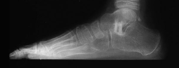 This is helpful to assess possible lateral impingement at the subtalar joint and subfibular impingement.
  If, on a simulated AP weight-bearing view with the eversion stress, there is adduction at the talonavicular joint or there is almost no eversion in the hindfoot, the foot is overcorrected. FootEducation LLC The incision was carried down through the skin and subcutaneous tissue with a #15 blade knife. WebA lateral column lengthening is performed typically to correct the forefoot abduction aspect of the deformity. For the first 6-10 weeks, the patient is either non-weight bearing or limited weight bearing through the heel, until the bone graft has healed. That situation will lead to an unsatisfied patient with lateral weight bearing. For the next 4-6 weeks (assuming the bone graft has healed), the patient can weight bear as tolerated in a cast boot. Ritchie (ankle-level hinged brace with plantar orthotic component). Our surgeon is a foot and ankle specialist, and he did an Evans procedure (lateral column lengthening) on a patient, and I am not sure how to code this.-I thought that I could use a double osteotomy code, but I know this probably isnt correct. WebLateral column lengthening has been used successfully in the treatment of stage II adult-acquired pes planovalgus deformity. It covers the incision, the desired outcome, the osteotomy and then two different methods of fixation. With [], Check Global Package and Graft Terminology Before Finalizing Your ACL Claim, Some extra services mean more codes but many dont. 26.5 Preoperative Preparation and Patient Positioning Measure the depth of the K-wire when it has reached the medial cortex. That situation will lead to an unsatisfied patient with lateral weight bearing. 26.1 Indications and Pathology is then placed.
26.3). In order to correct the calcaneal deformity, the tendon has to be detached from the calcaneus (as was done in this case), and the boney prominence, +/- any spurs, must be removed.  The interval between the facets can usually be identified bluntly with an elevator. Success with an LCL and cotton osteotomy is defined by achieving the right amount of correction with good alignment of the talonavicular and subtalar joints, resolving subtalar impingement and abduction of the talonavicular joint yet avoiding an overly stiff adducted/lateral weight-bearing foot. The op report describes a smaller incision than the desk reference and there is no mention of separation of the gastrocnemius from the soleus. WebLateral column lengthening procedures, either an Evans-type procedure or a calcaneocuboid distraction arthrodesis, clearly have a role to play in the management of a pes planovalgus foot deformity, as is evident from clinical outcome studies. WebLateral column lengthening with VariAx plate. This will give the surgeon a good idea of the depth for the saw blade cut. If there is any space on either side between the graft and native bone, rotate or trim the graft slightly to achieve excellent apposition along the lateral and dorsal aspects of the osteotomy. In adults and some children/adolescents, this prominence to which the Achilles Tendon attaches can lead to Calcaneal Bursitis and distal Achilles Tendinitis. Full recovery from flatfoot surgery Frisco, TX 75034 Best position is toes pointing to the ceiling with the foot at rest. for professional medical advice, diagnoses or treatments. Clinically, there should be near-normal eversion motion remaining, but mild stiffness is acceptable (Fig. For this procedure, you should report 28300 (Osteotomy; calcaneus [e.g., Dwyer or Chambers type procedure], with or without internal fixation). I agree that 28260 and 28300 do not appear to be appropriate for what was done. Borderline X-ray findings of one or two, but the patient has excessive pronation (eversion and abduction) seen clinically by a severe flatfoot with sag in the arch just distal to the ankle but not at the level of the tarsometatarsal or naviculocuneiform joints. Dissect at the midportion of the incision to find the floor of the sinus tarsi, taking care to avoid and stay above the peroneal tendons and sural nerve. 26.5). For a better experience, please enable JavaScript in your browser before proceeding. With the graft in place and pinned, confirm that the amount of correction is appropriate and that both clinical inspection and fluoroscopic views show good apposition of the graft to the native bone. If available, obtain a standing computed tomography (CT) scan in cases of severe deformity. I agree that 28260 and 28300 do not appear to be appropriate for what was done. For this procedure, you should report 28300 (, 5 Changes Will Update Your Sedation, Biomechanical Device, and Fracture Treatment Services, Plus: Dont miss these biopsy, fluoroscopic guidance revisions. document.getElementById( "ak_js_2" ).setAttribute( "value", ( new Date() ).getTime() ); The Relief Institute
  The successful patient has near-normal eversion motion remaining in the hindfoot, and good alignment of the heel. This should be explained to the patient. Spreading the cut bone apart with a bone or metal wedge helps recreate an arch. 12 weeks, patients usually can transition to wearing a shoe. Question:Our surgeon is a foot and ankle specialist, and he did an Evans procedure (lateral column lengthening) on a patient, and I am not sure how to code this.-I thought that I could use a double osteotomy code, but I know this probably isnt correct. Borderline X-ray findings of one or two, but the patient has excessive pronation (eversion and abduction) seen clinically by a severe flatfoot with sag in the arch just distal to the ankle but not at the level of the tarsometatarsal or naviculocuneiform joints. The bone graft is inserted in the joint, which serves as a joint fusion while also lengthening the lateral column. Weaken the medial cortex so that the osteotomy can be hinged open with an osteotome (Fig. WebLateral Column Lengthening In this procedure, the calcaneus bone is cut on the outside of the foot and "lengthened" to help correct the foot deformity. Afoot and ankle orthopaedic surgeonshould Activities such as walking, biking, driving, and even golfing are well tolerated. This is typically done by inserting either a cadaver bone or a metal wedge into the cut bone to lengthen it. of the foot, it is not a primary goal of treatment. Fill out the form below and we will call you back. Radiographically, the abduction should be corrected so that there is a normal amount of uncoverage of the talar head (30% or less), and no adduction of the talonavicular joint. This video demonstrates a lateral column lengthening. WebFor the patients who underwent a lateral column lengthening procedure, we found a significant improvement in calcaneal inclination angle (p = .001) and greater correction in talar declination angle, cuboid abduction angle, and talocalcaneal angle when compared with the control group. Experiences with VariAx 2 . The interval between the facets can usually be identified bluntly with an elevator. Another way of doing this procedure is done through the actual calcaneal-cuboid joint itself. The content is not intended to substitute WebLateral column lengthening with VariAx plate. Arizona brace. Fix the osteotomy with two longitudinal 3.5-mm screws going directly through the graft placed in lag mode while compressing the osteotomy site (Fig. WebLateral column lengthening with calcaneocuboid fusion, which lengthens the lateral column of the foot and prevents calcaneocuboid arthritis, was investigated in a cadaver model to determine the remaining range of motion in the talonavicular and subtalar joints. Only gold members can continue reading. Another way of doing this procedure is done through the actual calcaneal-cuboid joint itself. About 75% of the recovery occurs within the first 5-6 months. Clinically, there should be near-normal eversion motion remaining, but mild stiffness is acceptable (Fig. With the thin oscillating saw, make the osteotomy perpendicular to plantar aspect of the foot just distal to the K-wire into the medial cortex. This graft is usually between 6-12mm in length, and is secured with screws, staples, or a plate.
  For the next 4-6 weeks (assuming the bone graft has healed), the patient can weight bear as tolerated in acast boot. Indication for this procedure is excessive eversion/abduction of the midfoot with collapse of the arch as evidenced by one of the following: Forty percent or more talonavicular uncoverage on a standing AP X-ray of the foot. WebLateral column lengthening with VariAx plate. Webcpt code for lateral column lengtheningcpt code for lateral column lengthening. It may not display this or other websites correctly. By extending the length of the calcaneus at the location of the talonavicular joint, the talonavicular joint can be rotated from an abducted to neutral alignment. (214) 571-4581. It seems to be closest to either 28304 or 28305. 26.4 Key Principles of the Surgical Procedure The most common amounts of LCL are in the range of 6 to 8 mm. The depth of the saw cut can be marked on the saw with a marking pen. These complications often can be prevented with proper wound care and rehabilitation. pressure during standing and walking. It covers the incision, the desired outcome, the osteotomy and then two different methods of fixation. If available, obtain a standing computed tomography (CT) scan in cases of severe deformity. A flexor digitorum longus tendon transfer is usually performed in combination with the osteotomies in adult acquired flatfoot deformity with associated PTT pathology. For a better experience, please enable JavaScript in your browser before proceeding. Mobilize the peroneal tendons so that they can be retracted with a Bennett retractor to allow a saw cut into the lateral aspect of the anterior calcaneus. Judge the abduction of the talonavicular joint on the AP foot X-ray and the plantar sag at the talonavicular joint on the lateral X-ray. Use standing X-rays preoperatively, with the patient allowing the arch to collapse. Alternative fixation is with a lateral low-profile claw-type plate to provide compression. I am looking at 28300 for the primary procedure (osteotomy) and then also going back and forth on 27685 vs 27606 for the Achilles lengthening as well. When this is achieved, place a pin from the anterior calcaneus across the graft and into the posterior calcaneus. close the osteotomy site down and hold with 1.6mm wire or a staple, repair with side to side interrpted 2-0 nonabsorbable sutures after lengthening tendon to appropriate tension, plicate capsule with size 1 absorbable or non-absorbable suture in an interrupted or figure-8 fashion, advance the proximal slip of the tibialis posterior approximately 5 to 7 mm through a slit in the distal slump of the tendon using a pulvertaft weave with an absorbable suture material, alternatively sew tendon in a side to side fashion with 2.0 interrupted sutures, 2-0 or 3-0 absorbable suture for subcutaneous tissue, 3-0 absorbable, undyed running monofilament for medial incision, 3-0 non-absorbable mattress sutures are used for the lateral, calcaneal incision, place in a bivalved non weightbearing short cast, gait training for strict non weight bearing on operative side, do not get cast wet or insert anything into cast, return to OR for bone grafting and internal fixation with screw or plate, evaluate lab work cbc with diff, sed rate, crp, treat with dressing changes and oral antibiotics when appropriate, return to OR for irrigation, debridement, and IV antibiotices when necessary, treat with early mobilization and physical therapy for desensitization, refer to Pain Management if patient does not respond quickly to mobilization and desensitization. An osteotomy (bone cut) of the calcaneus is performed right before the calcaneal-cuboid joint, which is then spread about 7-10 mm so that the bone graft can be inserted, in order to lengthen the column (Figure 2). AlloSync Evans Wedge, 18 mm x 18 mm x 6.5 mm, AlloSync Evans Wedge, 18 mm x 18 mm x 8 mm, AlloSync Evans Wedge, 18 mm x 18 mm x 10 mm, AlloSync Evans Wedge, 18 mm x 18 mm x 12 mm, AlloSync Evans Wedge, 20 mm x 20 mm x 6.5 mm, AlloSync Evans Wedge, 20 mm x 20 mm x 8 mm, AlloSync Evans Wedge, 20 mm x 20 mm x 10 mm, AlloSync Evans Wedge, 20 mm x 20 mm x 12 mm, AlloSync Evans Wedge, 22 mm x 22 mm x 6.5 mm, AlloSync Evans Wedge, 22 mm x 22 mm x 8 mm, AlloSync Evans Wedge, 22 mm x 22 mm x 10 mm, AlloSync Evans Wedge, 22 mm x 22 mm x 12 mm, Plate, Low Profile, Cotton, Titanium, Flat, Plate, Low Profile, Cotton, Titanium, 2 mm, Plate, Low Profile, Cotton, Titanium, 4 mm, Plate, Low Profile, Cotton, Titanium, 6 mm, Plate, Low Profile, Cotton, Titanium, 8 mm. Any medical and health-related information presented on this website is general in nature. 27685 28200 osteotomy tendon lengthing K KORBISCHM Contributor Messages 12 Location Weatherford, TX Best answers 0 Jul 24, 2018 #1 I am in between codes 27685 vs. 28200. Fashion the graft according to the ideal amount of correction as shown by looking at the osteotomy held open to the desired amount.