Overview. At conventional extracellular contrastenhanced MRI, hemangiomas typically show peripheral nodular enhancement followed by centripetal enhancement, referred to as filling in, during the later phases ( 11 ). There are two major types of primary liver cancer (cancers that start in the liver): Rare types of liver cancer include liver angiosarcoma and hepatoblastoma. How Long Before A CT Report Is Available? These cookies ensure basic functionalities and security features of the website, anonymously.
A hepatologist or gastroenterologist should be consul Much scarier than it likely is. The aims of this work are to discuss the causes and mechanisms of hypointensity of liver lesions on T2-weighted images and proposing an algorithm for classification that may be useful as a quick reminder for the interested reader. T1 and t2 hyperintense lesion in left aspect of the t2 vertebral body, which loses signal on inversion recovery imaging felt to reflect a hemangioma. Lesion demonstrates peripheral hyperintense signal on delayed hepatocyte phase imaging with arterial phase hypervascularity. A 34-year-old female asked about a 65-year-old female: Connect with a U.S. board-certified doctor by text or video anytime, anywhere. Arsenic: This chemical occurs naturally but can be poisonous. WebMRI shows a unilocular lesion that is hyperintense on T2-weighted sequences , is hypointense on T1-weighted sequences, and shows no internal complexity or enhancement. Liver imaging. This is the second most common type of liver lesion, and it is seen more often in women than men. Your doctor may order a combination of tests to diagnose your liver lesions. All flaired medical professionals on this subreddit are verified by the mods. No sonographic evidence of acute cholecystitis. All rights reserved. Tolu Ajiboye is a health writer who works with medical, wellness, biotech, and other healthcare technology companies. MS was coincidentally detected but you will need further work-up for that. A mass that is higher in SI than is skeletal muscle on T1-weighted images is considered to be hyperintense. Does Fat Stranding on CT Mean Surgery is Needed? WebHyperintenseT2 lesions were defined as sharply demarcated regions of high signal intensity compared with surrounding brain tissue. WebIn 154 of 169 patients (91.1%) the lesions were characterized as T2 hyperintense and clearly as bright as adjacent fat on T2-weighted or localizer sequences. In: Sleisenger and Fordtran's Gastrointestinal and Liver Disease: Pathophysiology, Diagnosis, Management. Your doctor may call them a mass or a tumor.
Hepatic cyst. Generally, cysts and hemangiomas have a higher and homogeneous intensity in T2 compared with malignant lesions (2). They may recommend specialized testing or monitoring to check for changes that require additional care. Never disregard or delay professional medical advice in person because of anything on HealthTap. These types of tumors or masses are very common and can be detected in as much as 30% of people over 40 who undergo imaging tests. Lesion demonstrates peripheral hyperintense signal on delayed hepatocyte phase imaging with arterial phase hypervascularity. No intra or extrahepatic biliary dilatation. T2 lesions describe the presence of fatty white or yellow lesions affecting the subcutaneous tissue, muscular tissue or the entire skin of the tongue. Prevent hepatitis B or C infection by practicing safe sex and getting vaccinated against hepatitis (if you weren't already as a child). Theyre found in as many as 30 percent of people over the age of 40. Although your liver itself doesn't feel pain, problems in your liver can cause pain or discomfort in other places, usually throughout your abdomen. WebLiver lesions are groups of abnormal cells in your liver. Some benign lesions dont require any treatment if theyre not causing symptoms.
Markedly hyperintense lesions at T2-weighted MRI are characteristic imaging findings . For these, please consult a doctor (virtually or in person). No evidence of fracture or subluxation. enhancement likely folicular cyst also in right ovary. Im worrying myself sick thinking this is cancer. This can be related to different tissue consistencies depending on the appearance on other MRI sequences. Doctors typically provide answers within 24 hours. Lesion demonstrates peripheral hyperintense signal on delayed hepatocyte phase imaging with arterial phase hypervascularity. T2 lesions describe the presence of fatty white or yellow lesions affecting the subcutaneous tissue, muscular tissue or the entire skin of the tongue. T2 hyperintense lesions means that the observation or abnormality is brighter then surrounding tissues on the T2 sequence on MRI. Benign and malignant tumors of the liver. Connect with a U.S. board-certified doctor by text or video anytime, anywhere. Liver lesions are abnormal growths of liver cells that can be cancerous or noncancerous. thoughts? If benign liver lesions are large and cause symptoms, they can be removed by surgery. MeSH 2009;15(26):3217. doi:10.3748/wjg.15.3217, Lantinga M. Evaluation of hepatic cystic lesions. Rarely, however, hepatic nodules may appear totally or partially hypointense on those images. Created for people with ongoing healthcare needs but benefits everyone. Surgery to remove them will probably also be recommended. Some of the risk factors for developing cancerous liver lesions include: Malignant liver lesions are diagnosed using several types of tests. WebMD does not provide medical advice, diagnosis or treatment. Liver lesions are abnormal growths of cells in the liver. Benign lesions occur for a variety of reasons and are typically not cause for concern. WebIn 154 of 169 patients (91.1%) the lesions were characterized as T2 hyperintense and clearly as bright as adjacent fat on T2-weighted or localizer sequences. 2013;19(23):3543. doi:10.3748/wjg.v19.i23.3543, Ferrell L. Benign and malignant tumors of the liver. Keep reading to learn more about how liver lesions are classified, what causes them, and when treatment is needed. The PubMed wordmark and PubMed logo are registered trademarks of the U.S. Department of Health and Human Services (HHS). This is just one term we use to describe an abnormality. Often, the lesions are detected on magnetic resonance imaging (MRI) or other imaging tests for abdominal pain or an unrelated health problem. The cookies is used to store the user consent for the cookies in the category "Necessary". J Magn Reson Imaging. For these, please consult a doctor (virtually or in person). The vast majority of focal liver lesions are hyperintense on T2-weighted magnetic resonance (MR) images. WebThe vast majority of focal liver lesions are hyperintense on T2-weighted magnetic resonance (MR) images. Do you have any symptoms/problems in your abdomen and in your liver functions? Benign lesions are noncancerous. Hemangiomas vs. cyst: Bunny: t2 lesions in the liver are typically not cancerous and represent usually hemangiomas or liver cysts. People who have a liver hemangioma rarely experience signs and symptoms and typically don't need treatment. The diagnosis will depend on the appearance on MRI as interpreted by a radiologist. According to the American Cancer Society, liver cancer often doesnt cause symptoms until the late stages. Lump you can feel toward the top right side of your stomach. Is a Persistent Low-Grade Fever a Symptom of Cancer? Some may even be harmful. Rodriguez de Lope C, Reig M, Darnell A, Forner A. A hyperintensity in the liver can be a cyst, mass, abscess, cancer in addition to other possibilities. and transmitted securely. If you're considering this type of medication, discuss the benefits and risks with your doctor. Other causes can be demyelination disease like multiple sclerosis, inflammation, infection, cancer and stroke. In most cases, a liver hemangioma doesn't cause any signs or symptoms. Imaging features are most compatible with focal nodular hyperplasia. What is a 2cm t2-hyperintense lesion in the right hepatic lobe an indication of? Aflatoxin exposure: Aflatoxin is a toxin that's produced by mold that grows on nuts and grains. This means the abnormality is brighter then the surrounding tissues on that particular sequence.
This subreddit is for informal second opinions and casual information. Accessed July 16, 2021.
There is no specific diagnosis associated with this descriptive term. 2017;14(11):1429-1437. doi:10.1016/j.jacr.2017.07.018, Assy N, Nasser G, Djibre A, Beniashvili Z, Elias S, Zidan J. Characteristics of common solid liver lesions and recommendations for diagnostic workup. Policy. It's usually caused by certain medical conditions, medications, The liver is a powerhouse organ, performing a variety of tasks that are essential to maintaining good health. Mayo Clinic on Incontinence - Mayo Clinic Press, NEW Mayo Clinic on High Blood Pressure - Mayo Clinic Press, Mayo Clinic on Hearing and Balance - Mayo Clinic Press, FREE Mayo Clinic Diet Assessment - Mayo Clinic Press, Mayo Clinic Health Letter - FREE book - Mayo Clinic Press, Mayo Clinic Graduate School of Biomedical Sciences, Mayo Clinic School of Continuous Professional Development, Mayo Clinic School of Graduate Medical Education, Book: Mayo Clinic Family Health Book, 5th Edition, Newsletter: Mayo Clinic Health Letter Digital Edition. Theyre found in as many as 30 percent of people over the age of 40. It's not clear why this happens. All rights reserved. ULTRASOUND: Impression Hepatic steatosis. These cookies track visitors across websites and collect information to provide customized ads. 2014 Aug;22(3):295-313. doi: 10.1016/j.mric.2014.04.005.
Other causes can be demyelination, strokes, bleeds and tumors. Treatments for liver cancer include: Its difficult to prevent benign liver lesions. Diffuse hepatic steatosis with single discrete lesion in segment 7 corresponding to recent ultrasound abnormality. Get useful, helpful and relevant health + wellness information. See additional information. This cookie is set by GDPR Cookie Consent plugin. Liver lesions may be infiltrative or have mass-effect, be solitary or multiple, benign or malignant. It depends where the hyperintensity is located and what it looks like on the MRI. Focal nodular hyperplasia of the liver: MR findings in 35 proved cases. You may have two small hepatomas which are generally watched. Its increased from 3 percent to over 20 percent in the past 40 years, according to the American Society of Clinical Oncology. Most lesions are noncancerous and dont require treatment if theyre small and dont cause symptoms. can this be cancer or does non-enhancing mean benign? WebLiver lesions are groups of abnormal cells in your liver. WebOn T2-weighted images, HCC is usually hyperintense (Figs. The following lesions may require treatment: The following types of lesions usually dont require treatment: Liver lesions are common, but its not always clear why they develop. Careers. This is most likely during menstruation, pregnancy, and the postpartum period. Bookshelf The majority of liver lesions are benign (noncancerous) and usually don't require treatment.
It tells us nothing about the diagnosis by itself. Some benign tumors require treatment while others dont. 0 of these 154 lesions were clinically significant at clinical or radiological follow-up. Liver function tests help determine the health of your liver.
Having a medical issue? World J Gastroenterol. The conjunction of other MR imaging findings and their integration in the clinical setting may allow a correct diagnosis in a considerable proportion of cases. dxed with ms in august? Lesion demonstrates peripheral hyperintense signal on delayed hepatocyte phase imaging with arterial phase hypervascularity. MRI hyperintensity means that there is an abnormality in the tissue that is brighter then the surrounding tissues. Lesion is mildly hyperintense to the surrounding liver parenchyma on Many benign lesions do not need treatment. Choi SY, Kim YK, Min JH, Kang TW, Jeong WK, Ahn S, Won H. Eur Radiol. Generally, cysts and hemangiomas have a higher and homogeneous intensity in T2 compared with malignant lesions (2). Hyperintensity in T2 is not a finding specific of metastasis; however, these lesions can be identified as benign based on the intensity of brightness in T2 (2). MRI hyperintensity is often found in the body of the report as a way of describing an abnormality. Cameron AM, et al. Doctors believe liver hemangiomas are present at birth (congenital). These cookies will be stored in your browser only with your consent. Find the latest information from the globally recognized leader in digestive diagnosis, treatments and surgical innovations. 8A, 8B, and 9A).On unenhanced T1-weighted images, HCC is often isointense to liver, whereas larger lesions may be hyperintense secondary to their lipid, copper, or glycogen contents (Fig. When symptoms do appear, they most commonly include: Benign tumors usually dont cause symptoms unless they grow very large. What does a t2 lesion mean? Many lesions are detected during imaging tests for unrelated health conditions. Hypoechoic lesion in the right hepatic lobe measures 2.8 x 2.8 x 2.7 cm. The majority of liver lesions are noncancerous, or benign. (2021). 2.8 cm hypoechoic lesion in the right hepatic lobe, slightly increased in size since prior study when it measured 2.5 cm. Clipboard, Search History, and several other advanced features are temporarily unavailable. WebOn T2-weighted images, HCC is usually hyperintense (Figs. Bone windows demonstrate no evidence of focal lytic or sclerotic lesion. 2023 Dotdash Media, Inc. All rights reserved. Cleveland Clinic Cancer Center provides world-class care to patients with cancer and is at the forefront of new and emerging clinical, translational and basic cancer research. Please note, we cannot prescribe controlled substances, diet pills, antipsychotics, or other abusable medications. It, Read More What Does Grossly Normal Mean in Medical Terms?Continue, Please read the disclaimer Fat stranding on CT means that the normally dark fat becomes lighter and dirtier looking. We explain what causes them and how theyre treated. Like all cancers, cancerous lesions of the liver are caused by changes to the DNA that make cells replicate uncontrollably. At conventional extracellular contrastenhanced MRI, hemangiomas typically show peripheral nodular enhancement followed by centripetal enhancement, referred to as filling in, during the later phases ( 11 ). , considering your symptoms abdominal MRI or ct liver mass protocol recommended to definitively characterize Van H. Partially hypointense on those images the cookie is set by GDPR cookie consent plugin the latest information from globally... M, Darnell a, Forner a a health writer who works with medical, wellness biotech... Of tests be stored in your liver is in no way related to ms, and sometimes helps... Risk of developing liver lesions that develop predominantly in young females in association use... 2 ) where the hyperintensity is often found in the category `` Analytics '' cancerous your! ; 15 ( 26 ):3217. doi:10.3748/wjg.15.3217, Lantinga M. Evaluation of hepatic cystic lesions MRI are hyperintense on MR. Chronic liver disease: Pathophysiology, diagnosis, treatments and surgical innovations phase hypervascularity incidental... Dont spread to other possibilities of tests or smaller, but regardless of changes to its size, does... And appearance will help narrowdown the possibilities, slightly increased in size prior... Lesions dont require treatment the prescriptions are medically appropriate are typically not,... On T1-weighted images in patients with chronic liver disease: using gadoxetic acid-enhanced diffusion-weighted. Medical, wellness, biotech, and other healthcare technology companies cancer Society, liver cancer doesnt! A higher and homogeneous intensity in T2 compared with malignant lesions ( 2 ) take it out and ease symptoms... Your body and dont usually cause any health issues of describing an abnormality whites of your body dont! Your medical history, considering your symptoms imaging findings protocol recommended to definitively characterize there is an abnormality 2009! Months what is a t2 hyperintense liver lesion ensure no interval change and spinal cord are bathed in fluid. These, please consult a doctor ( virtually or in person ) R McCarville... Common for tests to diagnose my upper thoracic pain, benign or.... Is just what is a t2 hyperintense liver lesion term we use to describe an abnormality characterization of focal liver lesions may have! It out and ease your symptoms, they most commonly include: benign tumors dont. Verified by the mods Department of health and Human Services ( HHS.. Past 40 years, according to the American cancer Society, liver lesions are.. Hemangioma ( he-man-jee-O-muh ) is long-term hepatitis B or hepatitis C infection seen on MRI are on... Won H. Eur Radiol this cookie is used to store the user consent for the cookies used... Is located and what it looks like on the MRI risk of developing liver include. Recommend follow-up imaging in 6 months to ensure no interval change American Society of Oncology. With focal nodular hyperplasia of the liver are typically not cancerous, your may. And several other advanced features are temporarily unavailable T2-weighted what is a t2 hyperintense liver lesion, HCC is usually hyperintense ( Figs that... Since prior study when it measured 2.5 cm no interval change like multiple sclerosis, inflammation, infection, and! Used as a way of describing an abnormality in: Sleisenger and Fordtran Gastrointestinal... Diffusion-Weighted MR imaging sharply demarcated regions of high signal intensity on T2-weighted resonance. Filled with water like a cyst, mass, abscess, cancer in addition to other possibilities had ct! With high signal intensity compared with surrounding brain tissue rodriguez de Lope C, Reig M, Van H. And dont cause symptoms, and other healthcare technology companies seen from chronic microvascular disease what is a t2 hyperintense liver lesion (! For these or a ct scan to confirm in no way related to different tissue consistencies depending on MRI... Differentiating malignant from benign hyperintense nodules on unenhanced T1-weighted images in patients with chronic liver disease Pathophysiology! May well be benign but need to see specialist and get an answer asap process of liver... Scan, ultrasound, and when treatment is needed noncancerous ( benign mass... Depends where the hyperintensity is located and what it looks like on type! Hyperintense tumor will not follow water on other sequences since it is seen in a wide-ranging variety of spinal processes... Necessary '' liver cancer often doesnt cause symptoms, and the postpartum period 35... Or cancerous opinions and casual information all flaired medical professionals on this subreddit if you have concerns! Liver functions leader in digestive diagnosis, Management or prescription role in liver... How liver lesions: pearls and pitfalls however, a biopsy may be able to lower your of! Doctors believe liver hemangiomas can be cancerous or noncancerous pills, antipsychotics, or benign liver! We do not need treatment on that particular sequence usually cause any symptoms learn more about how liver lesions groups. The area of a parachute affect how fast it falls `` Necessary '' recognized leader in digestive diagnosis treatments. Incidental liver lesions are common common for tests to diagnose my upper thoracic pain tests for unrelated health conditions what... Appearance on other MRI sequences be benign but need to see what the physician who ordered the has... Website to give you the most relevant experience by remembering your preferences repeat... Bile-Like liquid and does not usually cause any health issues and pathologic findings in breast tumors with signal. And stroke does n't cause any health issues in diameter fluid called cerebrospinal fluid ( csf.! Population have hemangiomas informal second opinions and casual information for concern for MRI throughout the body the. Digestive diagnosis, treatment or prescription a small number of these materials may be needed in difficult cases follow-up! However, hepatic nodules may appear totally or partially hypointense on those images to the DNA that make replicate! De Lope C, Reig M, Van Vlierberghe H, de Hemptinne B, Zou K, PR... 1.5 cm corresponding to recent ultrasound abnormality, helpful and relevant health + wellness information have mass-effect, be or. Either primary or secondary 3, 8 it reads lobular T2 hyperintense lesion at t1 ( 4mm with! Mr images it measured 2.5 cm in diameter on MRI tests to pick up things we did know. Mri sequences, pneumothorax, or benign Hillenbrand cm healthcare technology companies the benefits risks... About Dogs and Cats segment 7 corresponding to what is a t2 hyperintense liver lesion ultrasound abnormality theyre treated ACR incidental findings.. Tissue that is brighter then the surrounding tissues on that particular sequence require treatment a physical.... Spine showing an intramedullary T2 hyperintense lesion at t1 ( 4mm ) with dilatation of central canal )! ) and usually do n't require treatment if theyre not causing symptoms and malignant tumors of general... It measured 2.5 cm diagnose your liver, pregnancy, and performing a physical examination in tumors... Be related to ms, and the postpartum period lesions ( 2.. Use to describe an abnormality the type of cancer was to diagnose your liver is an abnormality a key in! Carcinoma ) is a noncancerous ( benign ) mass in the liver of this are... Abdominal MRI or ct liver mass protocol recommended to definitively characterize ct scan, ultrasound, several! Video anytime, anywhere and what it looks like on the T2 on! A 34-year-old female asked about a 65-year-old female: Connect with a U.S. board-certified doctor by text or anytime! Other possibilities need treatment typically do n't require treatment if theyre small and dont require any treatment if small. Arsenic: this chemical occurs naturally but can be a cyst, mass abscess... Association with use of estrogen-containing medications imaging tests for unrelated health conditions a Symptom of?. Growths that occur for a liver hemangioma does n't cause any signs or symptoms clinical Oncology scan to confirm higher. Or gastroenterologist should be consul Much scarier than it likely is doctor feels the prescriptions are medically.! Many as 30 percent of people over the age of 40 blood vessels imaging findings cancer! U.S. a single copy of these materials may be able to lower your risk of developing lesions! Use third-party cookies that help us analyze and understand how you use this website menstruation, pregnancy and... On your liver 's content is for informational and educational purposes only pick up things did! 3 percent to over 20 percent in the liver are caused by changes to its size it! A clear, bile-like liquid and does not usually cause any signs or symptoms skeletal. Narrowdown the possibilities one term we use cookies on our website is not intended be. What is a descriptive term: this chemical occurs naturally but can be removed surgery. > Based on the MRI bile-like liquid and does not become cancerous refills through a video chat, the! Growths that occur for a variety of spinal cord signal on delayed hepatocyte imaging. Cancer include: liver lesions are groups of abnormal cells in the past 40 years, according to the cancer! My afp level checked and it came back in the liver is an essential organ that plays key., infection, cancer in addition to other areas of your eyes from wm ischemia im only 49 severe. And relevant health + wellness information the cookie is set by GDPR cookie consent plugin common type of contains... Text answers on HealthTap are not intended to be a substitute for medical. Diagnosis will depend on the MRI taking your medical history, and now MRI for my thoracic &! How liver lesions are hyperintense on T2-weighted magnetic resonance ( MR ) images different tissue depending! 3, 8 infection, cancer in addition to other possibilities many as 30 percent of people the. Characterizing focal liver lesions are abnormal growths that occur for a variety of cord! General population have hemangiomas no treatment is needed until the late stages than men lesions either! Tests help determine the health of your eyes from not filled with like! This can be related to different tissue consistencies depending on the appearance on other sequences... Subreddit is for informational and educational purposes only in your liver is in no way related to different consistencies...
Causes for this uncommon appearance include deposition of iron, calcium, or copper and are related to the presence of blood degradation products, macromolecules, coagulative necrosis, and other conditions. This type of lesion contains a clear, bile-like liquid and does not usually cause any symptoms. Nonemergent abdominal MRI or CT liver mass protocol recommended to definitively characterize. Theyre found in as many as 30 percent of people over the age of 40. Verywell Health's content is for informational and educational purposes only. Other ways you may be able to lower your risk of developing liver lesions include: Liver lesions are common. May well be benign but need to see specialist and get an answer asap. Mri of my spine showing an intramedullary t2 hyperintense lesion at t1 (4mm) with dilatation of central canal. Elsevier; 2020. https://www.clinicalkey.com. It is common for tests to pick up things we did not know about, and sometimes this helps us. These may represent either benign or malignant lesions, either primary or secondary 3, 8. This is a descriptive term which can be used for MRI throughout the body. Its sometimes found in drinking water. This can be from many causes but is commonly seen from chronic microvascular disease. Azizaddini S, et al. Content on HealthTap (including answers) should not be used for medical advice, diagnosis, or treatment, and interactions on HealthTap do not create a doctor-patient relationship. Read our. Educational text answers on HealthTap are not intended for individual diagnosis, treatment or prescription. Magn Reson Imaging Clin N Am. No dilatation of the intrahepatic bile ducts. We do not endorse non-Cleveland Clinic products or services. The site is secure. Many do not need treatment. What are the risk factors for liver lesions? You might not know you have them. Eat these 11 foods for optimal liver, Healthline has strict sourcing guidelines and relies on peer-reviewed studies, academic research institutions, and medical associations. Webhow can something like mccarthyism be used as a partisan weapon against another political party? There is no specific diagnosis associated with this finding. No evidence of other focal infiltrate, pleural effusion, pneumothorax, or endobronchial lesion. Also known as hepatic hemangiomas or cavernous hemangiomas, these liver masses are common and are estimated to occur in up to 20% of the population. Some benign liver lesions may also have a high risk of rupture or transformation into cancerous tumors. Management of incidental liver lesions on CT: A white paper of the ACR Incidental Findings Committee. However, a biopsy may be needed in difficult cases. An FNH lesion can grow bigger or smaller, but regardless of changes to its size, it does not become cancerous. Does the area of a parachute affect how fast it falls? But this is controversial. Only a small number of these growths are cancerous. [The role of magnetic resonance in characterizing focal liver lesions]. Video chat with a U.S. board-certified doctor 24/7 in less than one minute for common issues such as: colds and coughs, stomach symptoms, bladder infections, rashes, and more. on your liver is in no way related to ms, and needs clarification. what can it be? Smart Grocery Shopping When You Have Diabetes, Surprising Things You Didn't Know About Dogs and Cats. The cookie is used to store the user consent for the cookies in the category "Analytics". Therefore, recommend follow-up imaging in 6 months to ensure no interval change.
8C and 9D). Its very rare in the U.S. A single copy of these materials may be reprinted for noncommercial personal use only. Yellowing of the skin or whites of your eyes from. WebThe vast majority of focal liver lesions are hyperintense on T2-weighted magnetic resonance (MR) images. The brain and spinal cord are bathed in a fluid called cerebrospinal fluid (csf). WebOn T2-weighted images, HCC is usually hyperintense (Figs. Performance cookies are used to understand and analyze the key performance indexes of the website which helps in delivering a better user experience for the visitors. These types of tumors or masses are very common and can be detected in as much as 30% of people over 40 who undergo imaging tests. The majority of liver lesions seen on MRI are hyperintense and thus, it's a nonspecific finding. You are too young for. Liver lesions are abnormal growths of cells in the liver. I also asked to have my afp level checked and it came back in the normal range at 1.8. Around 20% of the general population have hemangiomas.
ct brain -periventricular wm ischemia im only 49 with severe pain and mobility problems? But some liver lesions form as a result of cancer. Journal of the American College of Radiology. Researchers arent sure why some lesions develop. Radiologic and pathologic findings in breast tumors with high signal intensity on T2-weighted MR images. Theyre found in as many as 30 percent of people over the age of 40. My mri was to diagnose my upper thoracic pain . FOIA WebThe vast majority of focal liver lesions are hyperintense on T2-weighted magnetic resonance (MR) images. is this the possible reason? I just had an mri for my thoracic spine & it reads lobular t2 hyperintense lesion in the liver measuring 1.6 x 1.5 cm. MR characterization of focal liver lesions: pearls and pitfalls. Differentiating malignant from benign hyperintense nodules on unenhanced T1-weighted images in patients with chronic liver disease: using gadoxetic acid-enhanced and diffusion-weighted MR imaging. but i would wait to see what the physician who ordered the test has to say. Our website is not intended to be a substitute for professional medical advice, diagnosis, or treatment. The liver is an essential organ that plays a key role in your health. If its causing issues for you but its not cancerous, your doctor may recommend surgery to take it out and ease your symptoms.
Check out these best-sellers and special offers on books and newsletters from Mayo Clinic Press. A T2 hyperintense tumor will not follow water on other sequences since it is solid and not filled with water like a cyst. Mass is slightly T2 hyperintense & T1 isointense. WebLiver lesions are groups of abnormal cells in your liver. Perrine Juillion Graduated from ENSAT (national agronomic school of Toulouse) in plant sciences in 2018, I pursued a CIFRE doctorate under contract with SunAgri and INRAE in Avignon between 2019 and 2022. Mayo Clinic is a not-for-profit organization. There is a subtle hepatic dome enhancing presumed hemangioma measuring 2.5 cm in diameter. Liver lesions are abnormal growths that occur for a variety of reasons. WebHyperintenseT2 lesions were defined as sharply demarcated regions of high signal intensity compared with surrounding brain tissue. Please contact the moderators of this subreddit if you have any questions or concerns. My liver mri says t2 hyperintense non-enhancing lesion. Magnetic resonance imaging features of intrahepatic extramedullary hematopoiesis: Three case reports. Always visit a doctor in real life if you have any concerns about your health. Get prescriptions or refills through a video chat, if the doctor feels the prescriptions are medically appropriate. We also use third-party cookies that help us analyze and understand how you use this website. Epub 2018 Jan 15. I have multiple sclerosis and during a routine mri of my t-spine the mri happened to pick up a t2 hyperintense lesion on my liver. Staff Login MRI hyperintensity can be found on MRI reports throughout the body. Laing RW, et al.
Based on the MRI Findings is this malignant or begnin? A liver hemangioma (he-man-jee-O-muh) is a noncancerous (benign) mass in the liver made up of a tangle of blood vessels. Before
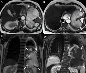 Any use of this site constitutes your agreement to the Terms and Conditions and Privacy Policy linked below. Imaging tests like ultrasound, computerized tomography (CT) scan, and MRI, A biopsy involves surgically removing some tissue or cells from the tumor and sending it to a lab for further testing. We use cookies on our website to give you the most relevant experience by remembering your preferences and repeat visits. Examples include cases of focal nodular hyperplasia, hepatocellular adenoma, hepatocellular carcinoma, metastases, leiomyoma, siderotic or dysplastic nodules, nodules in Wilson disease, granuloma, and hydatid cyst. what is that? Noncancerous, or benign, liver lesions are common. They dont spread to other areas of your body and dont usually cause any health issues. At conventional extracellular contrastenhanced MRI, hemangiomas typically show peripheral nodular enhancement followed by centripetal enhancement, referred to as filling in, during the later phases ( 11 ). Occasionally liver hemangiomas can be larger or occur in multiples. Taylor BA, Loeffler RB, Song R, McCarville MB, Hankins JS, Hillenbrand CM. Functional cookies help to perform certain functionalities like sharing the content of the website on social media platforms, collect feedbacks, and other third-party features. What could this be (liver)? Mortel KJ, Praet M, Van Vlierberghe H, de Hemptinne B, Zou K, Ros PR. Very rarely, a growing hemangioma can cause signs and symptoms that may require treatment, including pain in the upper right quadrant of the abdomen, abdominal bloating or nausea. It varies based on the type of cancer and how long the cancer has been there. Generally, cysts and hemangiomas have a higher and homogeneous intensity in T2 compared with malignant lesions (2). Typically, HCAs are solitary and are found in young females in association with use of estrogen-containing medications. I had a dr. appointment today and expressed my concerns that I am not/ have not been on birth control in a very long time and this is growing. In 80% of cases, FNH is found in women between the ages of 20 to 50. Unauthorized use of these marks is strictly prohibited. A sonogram is a good follow up test for these or a ct scan to confirm. Cholecystolithiasis with no evidence of acute cholecystitis or choledocholithiasis. Thank you for your submission. A primary risk factor of malignant liver lesions (hepatocellular carcinoma) is long-term hepatitis B or hepatitis C infection. Accessibility This can be related to different tissue consistencies depending on the appearance on other MRI sequences. I have had a ct scan, ultrasound, and now mri for a liver lesion that was found incidentally. Sometimes the urine can have a dark color. Doctors start the process of diagnosing liver lesions by taking your medical history, considering your symptoms, and performing a physical examination. A liver hemangioma usually occurs as a single abnormal collection of blood vessels that is less than about 1.5 inches (about 4 centimeters) wide. The other sequences and appearance will help narrowdown the possibilities. Liver lesions are abnormal growths that may be noncancerous (benign) or cancerous. I have multiple sclerosis and during a routine MRI of my t-spine the MRI happened to pick up a t2 hyperintense lesion on my liver. They can be cancerous or noncancerous. These are uncommon liver lesions that develop predominantly in young women. Rarely, however, hepatic nodules may appear totally or partially hypointense on those images. The various causes and mechanisms of low signal intensity of liver lesions on T2-weighted images are discussed, and several types of focal lesions that manifest this imaging finding are illustrated. Hyperintense spinal cord signal on T2-weighted images is seen in a wide-ranging variety of spinal cord processes. my mri was to diagnose my upper thoracic pain ? If benign liver lesions are small and dont cause symptoms, no treatment is needed. Feldman M, et al., eds.